

Key concepts:
- Isolation of Protoplasts
- Protoplasts isolation
- Purification of protoplasts
- Methods for protoplasts culture
- Viability of protoplasts
- Cell wall formation and cell division.
- Applications of protoplasts
Introduction
Protoplast refers to the cellular content excluding cell wall or can also be called as naked plant cell. It is described as, living matter which is cytoplasm and nucleoplasm enclosed in a plant cell membrane. The use of protoplasm in tissue culture technology offers fascinating option for the improvement of plants over the conventional breeding programs. How it is done and what prospects protoplast culture holds in itself, let us study and know about it in detail.
- Plant cells without the cell wall
- Naked spherical plant cells
- Functional plant cells
- Fragile
- Protoplasts can be regenerated into complete plants.
- Several applications in crop improvement

Protoplast Isolation
Protoplasts are isolated by:
- Mechanical Extraction
- Enzymatic Extraction
Mechanical Method
- Von Klercker (1892) isolated protoplasts, mechanically from an aquatic plant Stratiotes aloides. He first plasmolysed the leaf cells and then cut the cell walls to liberate the protoplasts.
- Ideal for isolating large and highly vacuolated cells of storage tissues such as onion bulb scales, radish root and beet root tissue.
- The cells are plasmolyzed in an iso osmotic solution resulting in the withdrawal of contents in the center of cell. Subsequently, the tissue is dissected and deplasmolyzed to release the preformed protoplasts.
- Plasmolysis is the process of shrinkage or contraction of the protoplasm of a plant cell as a result of loss of water from the cell.
- Plasmolysis is induced in the laboratory by immersing living cells in a strong salt or sugar solution to lose water from the cell.
- Once the protoplasm shrinks it can be separated out from the cell.
Limitations of mechanical isolation of protoplasts.
- It is restricted to certain tissues which have large vacuolated cells and is useful when there are side effects of cell wall degrading enzymes.
- Yield of protoplasts is generally very low. Protoplasts from less vacuolated and highly meristematic cells do not show good yield.
- The method is tedious and laborious.
- Viability of protoplasts is low.
Enzymatic method of separation of protoplasts
Many scientists have tried to modify or develop this technique, until later in 1960s Prof. Edward Cocking from Nottingham University, England, developed a sophisticated and novel approach for the large-scale production of higher plant protoplasts, using root tips of tomato seedlings. He used cellulase enzyme from the fungus Myrothecium, which involved the digestion of cellulosic cell walls and obtained protoplasts from the cells of the root tips.
The enzymatic separation of protoplasts can be done in two ways:
- One-step method, protoplasts are isolated directly from the tissues by using two enzymes, cellulase and pectinase, simultaneously.
- Two-step method (sequential method), cells are first isolated from tissues by using enzyme pectinase and to this cell suspension, cellulase is added to digest the cell wall and release protoplasts.
Enzymatic solution for these methods should be carefully prepared. The enzymatic solution should of course contain the enzymes but along with enzymes sugar should also be added. The sugar works as an osmoticum, which prevents the plasma membrane from rupturing, during cell wall digestion by enzymatic action. Along with sugar some salts and nutrients should also be used for providing stability and viability to the isolated protoplasts.
The enzymatic separation of protoplasts process involves four steps:
- Sterilization of leaves
- Peeling off the lower epidermis
- Incubation in enzyme solution and
- Isolation and purification of the protoplasts.
Sterilization of leaves
Healthy leaves are obtained from greenhouse grown plants and leaves are sterilized by 0.1% mercuric chloride solution followed by short treatment with 70% alcohol. The leaves are then rinsed three times with sterilized distilled water.
Peeling off the lower epidermis
After sterilization of the leaves, the lower epidermis is carefully removed and the strips are cut into small pieces.
Incubation in enzyme solution
- Mostly macerozyme and cellulase enzymes are used to obtain protoplasts in significant number.
- The pH of the enzyme solution is adjusted between 4.7 and 6.0.
- The osmotic concentration of enzyme mixture is elevated by adding 500- 800 m mol/l sorbitol or mannitol in order to prevent them from bursting. And to improve membrane stability usually CaCl2 (50-100mM) is also added to the enzyme solution.
- Along with this some salts like KCl and MgSO4 should be added as ionic osmoticum to maintain and improve protoplasts viability.
- Once the enzyme solution is ready, it is sterilized through a membrane filter and peeled leaf sections are placed in it.
- Petri-plates containing tissue and enzyme mixture are sealed with parafilm and incubated for 4-12 hours on a rocking shaker at 24-26 °C.
Isolation and Purification of Protoplasts
After incubation of plant tissues in enzyme solution, the solution is filtered through a wire or nylon mesh (50-100 µm) to remove debris like undigested cells, tissues, broken cells etc. The filtrate is transferred into screw capped small, sterilized centrifuge tubes and centrifuged at 100g. The protoplasts form a pellet at bottom while the small debris in the supernatant is carefully removed.
Advantages of Enzymatic Methods
- Easy to operate
- large quantities of protoplasts can be easily obtained.
- Viability percentage is higher.
- Osmotic shrinking is at minimum levels which was more in the mechanical method.
- Not restricted to specific tissues. Variety of plant tissues like leaves, roots, embryos, microspores etc. Suspension culture is good resource.
Purification of Protoplasts
- Prepare a sucrose gradient solution with the densest portion (highest percentage) at the bottom of the centrifuge tube. This can go from 40-45% sucrose from bottom to top.
- Layer the enzyme solution containing the protoplasts on top of the sucrose gradient.
- Centrifuge the tube. The protoplasts will migrate through the gradient until they reach the density zone that matches their own density.
- Carefully collect the protoplast band from the interface using a Pasteur pipette.
- Wash the collected protoplasts by resuspending in fresh sorbitol solution and repeating centrifugation 2-3 times. The protoplasts will float in 20% sucrose while debris pelletizes.
- Collect the floating clean protoplast layer with a pipette each time.
- Pool the washed protoplasts, count using a haemocytometer and dilute to the appropriate density for further culture or analysis.
Protoplast Viability
Once we obtain good yield of protoplasts, their viability can be checked by any of
the methods:
- Phase contrast microscopy: The viable protoplasts show cyclosis or cytoplasmic streaming which indicates active metabolism.
- Oxygen Uptake: Living protoplasts respire and respiratory metabolism can be measured by oxygen electrode.
- Evan’s Blue Test: The living protoplasts exclude evan’s blue with metabolism whereas dead protoplasts hold the dye inside them and are visualized blue under microscope.
- Fluroescein di acetate: The FDA is a neutral non fluorescent molecule which is taken up by living cells via passive diffusion and in the cytoplasm it is cleaved by enzyme esterases to give the fluorescent fluorescein which gives a green fluorescence under UV light.
Protoplast Culture
Since, the protoplasts are delicate as they are devoid of cell walls hence they have to be cultured with care. The commonly used technique for culturing protoplast is Bergmann’s Plating technique.
- Bergmann’s Plating Technique
- Micro Chamber Technique
- Micro drop method
Bergmann’s Plating Technique
This technique was proposed by Bergmann in 1960. In this technique the protoplasts are first suspended in a liquid medium with suitable osmoticum, at a density twice than the required density for cell plating. Then, equal volume of molten agar (30-35°C) is added to the medium containing protoplasts. The agar medium with protoplasts is spread out in a petridish. On cooling, the agar solidifies forming a thin jelly like layer. The petridishes are sealed with a parafilm and incubated at 25°C in dark or diffused light.
Micro Chamber Technique
This technique was used by Vasil and Hilderbrandt (1965) to raise tobacco plants from isolated cells. A drop of culture medium containing one or more protoplasts is put on a microscopic slide. On either side of this drop, two coverslips are kept. A third one is put over these two covers slips to shield the protoplasts suspensions. The edges of the coverslips are sealed with oil or grease and protoplasts are maintained under optimum culture conditions for growth.
Micro drop method
This method has been especially useful for culturing individual protoplasts. The technique was used by Kao (1977) to culture individual protoplasts. They used special Cuprak dishes which have two chambers, a small outer chamber and a large inner chamber. The large chamber carries numerous numbered microwells in which the protoplast suspension is transferred as microdroplets. The outer chamber is filled with sterile distilled water to maintain the humidity inside the dish. After covering it with the lid the dish is sealed with parafilm.
Protoplast Culture Requirements
- Culture medium
- Nutrients
- Osmotic pressure
- Plant Growth regulators
- Additional supplements
Protoplast Culture Conditions
Protoplast culture conditions, such as the use of liquid or semisolid medium, temperature and light, cell density, or the presence of nurse cultures, can have a significant effect on the division and microcallus formation potential of protoplasts.
- Liquid/Semi-solid medium
- Temperature and light
- Cell density
- Nurse culture
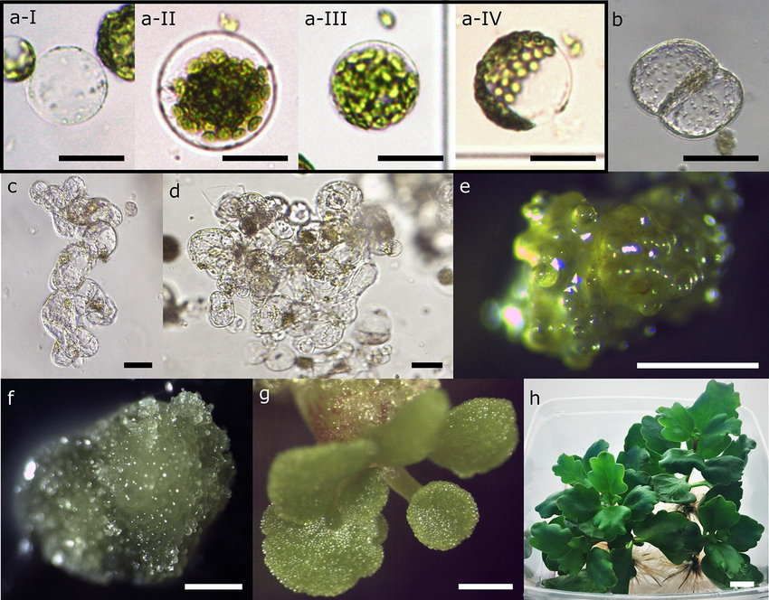
Cell wall formation
Under optimum conditions, during culture period, within 2-4 days, protoplasts lose their characteristic spherical shape. This indicates the formation of new wall. It is observed that mesophyll cells, callus and cell suspension protoplasts of most Solanaceous and many Brassica species form cell wall very quickly that is
within 24-40 h of culture. In contrast cereal protoplasts and mesophyll protoplasts of legume may require up to four days for cell wall regeneration. An even longer phase; 7 days or more is required for wall formation by protoplasts of the woody plants.
Cell Division and callus formation
There is a direct relationship between wall formation and cell division. Protoplasts which are not able to regenerate a proper wall fail to undergo normal mitosis. The protoplasts capable of dividing, undergo the first division within 2-7 days and form callus. On transferring callus to shoot induction medium, young shoot buds emerge which are then transferred to rooting medium for development of complete plantlets.
Several factors influence divisions in protoplast cultures:
- Nutritional requirements: Using optimized media like MS or B5 with appropriate growth hormones (auxins, cytokinins), reduced ammonium, and antioxidants.
- Osmoticum: Using mannitol or sorbitol for initial osmotic protection, then gradually reducing osmolarity over 7-10 days as cell walls regenerate.
- Plating density: Maintaining low initial density (100-500 protoplasts/ml) prevents early fusion of colonies.
- Physical treatment: Electroporation can trigger earlier cell divisions and improve efficiency.
- Storage conditions: Keeping cultures in low light or darkness initially, then transferring to light after cell wall regeneration. Maintaining 25-30°C temperature.
- Plant material: Using physiologically active donor tissues from controlled growth conditions. Axenic shoot cultures give high reproducibility. Explant source affects culture response.
Plant regeneration: First achieved in tobacco in 1971, now extended to diverse species including cereals, legumes, cotton, trees.
Applications of Protoplast Culture
- Plant breeding and crop improvement:
- Protoplast fusion to combine desirable traits from different species
- Generation of novel genetic variability
- Somaclonal variation for selection of variants
- Haploid production for breeding programs
- Genetic engineering by direct DNA uptake
- Totipotent, can regenerate cell walls and whole plants
- Can be used for genetic engineering and regeneration of genetic transformants.
- Somatic Hybridization: Fusion of protoplasts help to generate interspecific hybrids.
- Good experimental system to study plant-virus interrelationships.
- Induction of somatic embryos
Summary
Plant protoplasts provide a unique single cell system to underpin several aspects of modern biotechnology. Reliable procedures are available to isolate and culture protoplasts from a range of plants, including both monocotyledonous and dicotyledonous crops. Several parameters, particularly the source tissue, culture medium, and environmental factors, influence the ability of protoplasts and protoplast derived cells to express their totipotency and to develop fertile plants.
Nevertheless, isolated protoplasts are unique to a range of experimental procedures. In the context of plant genetic manipulation, somatic hybridization enables nuclear and cytoplasmic genomes to be combined, at the interspecific and intergeneric levels to overcome naturally occurring sexual incompatibility barriers. The protoplasts also provide systems for investigating most aspects of plant cell physiology and genetics, including proteomics.
Somatic Hybridization: Methods of Protoplast Fusion, Asymmetric and symmetric hybrids
Further reading
References
- Protoplast – Wikipedia
- Introduction to Plant Biotechnology, Third Edition, H.S. Chawla. ISBN 978-1-57808-636-8
- Tiew, Terence & Sheahan, Michael & Rose, Ray. (2015). Peroxisomes contribute to reactive oxygen species homeostasis and cell division induction in Arabidopsis protoplasts. Frontiers in Plant Science. 6. 10.3389/fpls.2015.00658. ↩︎
- Cui, Jin & Mackenzie, Kathryn & Eeckhaut, Tom & Müller, Renate & Lütken, Henrik. (2019). Protoplast isolation and culture from Kalanchoë species: optimization of plant growth regulator concentration for efficient callus production. Plant Cell, Tissue and Organ Culture (PCTOC). 138. 10.1007/s11240-019-01624-4. ↩︎